Left Hip Muscles Anatomy / Snapping Hip Orthoinfo Aaos : Skeletal muscle cells are multinucleate.
byAdmin•
0
Left Hip Muscles Anatomy / Snapping Hip Orthoinfo Aaos : Skeletal muscle cells are multinucleate.. The gluteal muscles work together to extend we're not suggesting you memorize gray's anatomy. Now that you watched the video, you. In clinical anatomy the thigh muscles are divided into three groups: The muscles of the hip and thigh keep your hip joints strong and mighty, allowing for a wide range of hip movements. Front view of the hip joint bones.
Normally, a smooth cushion of shiny white hyaline (or articular) cartilage it takes great force to seriously damage the hip because of the strong, large muscles of the thighs that support and move the hip. Numerous muscles and muscle groups surround the hip joint and play various roles in movements of the thigh. Now that you watched the video, you. How many of the 11 muscles involved in hip flexion can you name from memory? 1, tensor fasciae latae m.
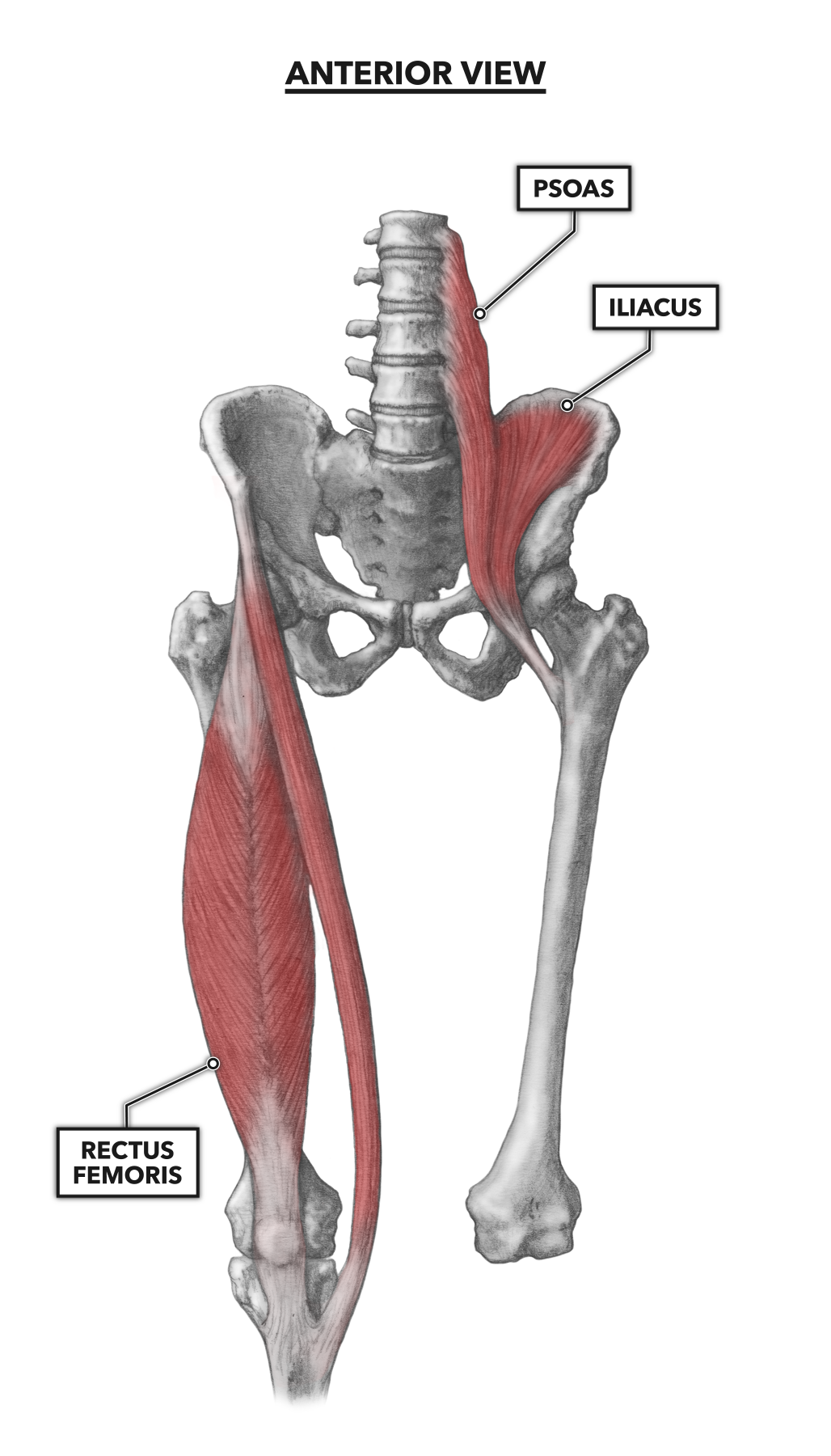
Muscles Of The Leg And Foot Classic Human Anatomy In Motion The Artist S Guide To The Dynamics Of Figure Drawing from doctorlib.info Move both the hip and knee quadriceps, hamstrings, deep '6' lateral rotators, gluteals, a… Hip joint muscles are divided into four groups according to their orientation and function. 1, tensor fasciae latae m. The iliopsoas muscle is a major hip flexor. Common action is external rotation. Learning the anatomy of your hip will better enable you to pinpoint your pain and work with your doctor to keep it from limiting your life. These muscles constitute the anatomical classification known as the medial compartment of the thigh. How many of the 11 muscles involved in hip flexion can you name from memory?
The anatomical basis of clinical practice (41st edition).
Individuals with obesity can have great difficulty maintaining abductor muscular function due to being overweight and possibly experiencing a decrease in muscle mass. The gluteal muscles work together to extend we're not suggesting you memorize gray's anatomy. In human anatomy, the muscles of the hip joint are those muscles that cause movement in the hip. Common action is external rotation. The hip and hip joint anatomical chart is a clean and clearly illustrated view on the human anatomy regarding the hip bones and muscles. Microscopic anatomy of skeletal muscle. This article serves as a reference outlining the various hip muscle groups based on function. In clinical anatomy the thigh muscles are divided into three groups: It's hard to remember them all! Now that you watched the video, you. We're just asking you to put your adrenaline on hold while we give you a rundown of your. 24.19 muscles of the hip, thigh, and gluteal region: Anatomical components of the hip and discuss the relevant.
24.19 muscles of the hip, thigh, and gluteal region: The anatomical basis of clinical practice (41st edition). The iliopsoas muscle is a major hip flexor. Almost all muscles cross at least one joint (moveable connection between two bones) and cause an action across that joint. Learning the anatomy of your hip will better enable you to pinpoint your pain and work with your doctor to keep it from limiting your life.
Muscles Of The Leg And Foot Classic Human Anatomy In Motion The Artist S Guide To The Dynamics Of Figure Drawing from doctorlib.info Most modern anatomists define 17 of these muscles, although some additional muscles may sometimes be considered. We're just asking you to put your adrenaline on hold while we give you a rundown of your. The hip is a complicated mechanism and therefore hip pain can originate in many different parts of the joint. Muscles that act on the lower limb cause movement at the hip, knee and foot joints. The gluteal muscles work together to extend we're not suggesting you memorize gray's anatomy. Muscles of hip and their action. This webpage presents the anatomical structures found on hip mri. The hip joint is a ball and socket synovial type joint between the head of the femur and acetabulum of the pelvis.
I pulled some muscles on left hip hiking.
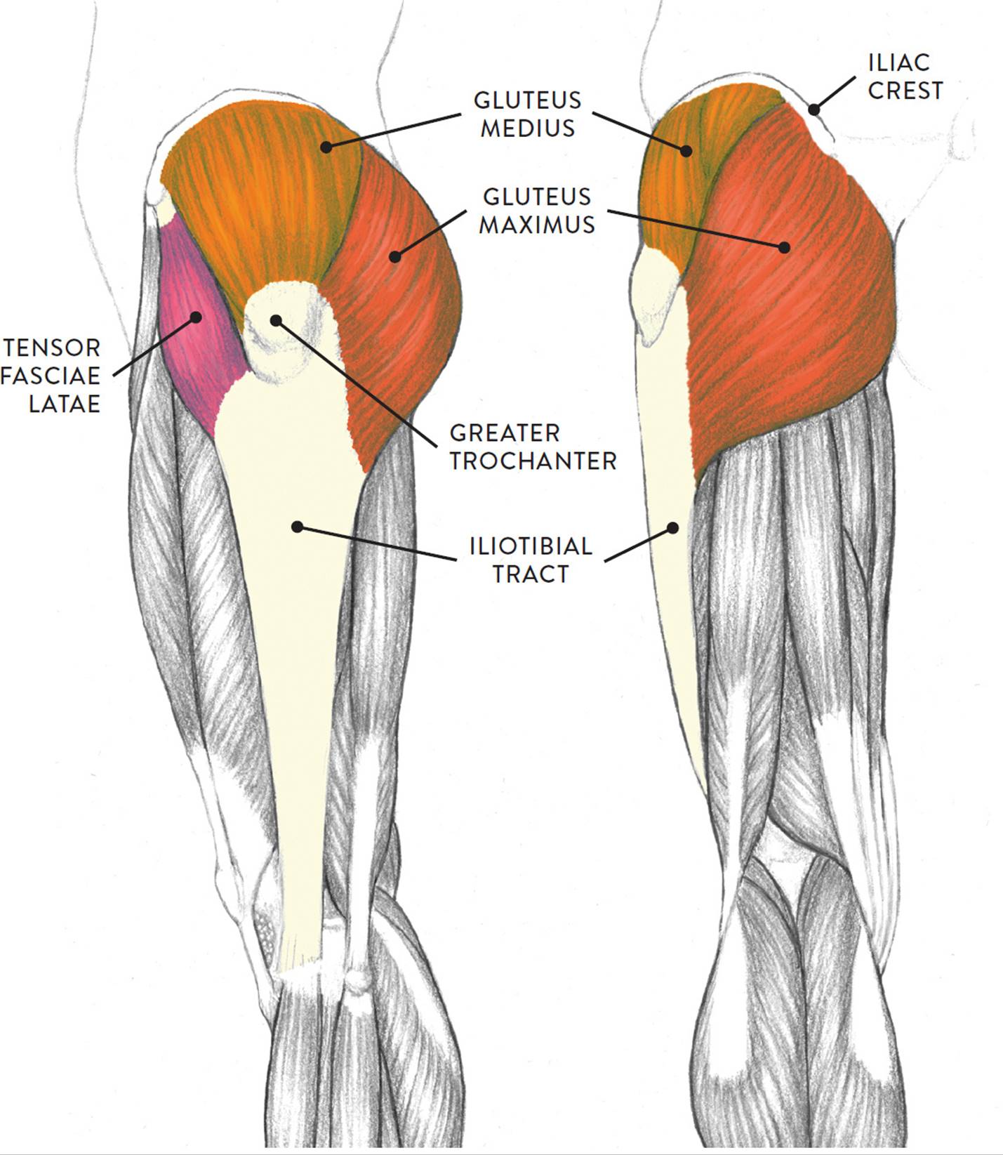
Rectus femoris forms the middle portion of the quadriceps. The muscles of the pelvis, hip and buttock anatomical chart shows how each muscle in this area of the body works with the others, and the various minor systems within the major ones. Learn their anatomy efficiently and easily using kenhub's muscle anatomy and reference charts! The iliotibial tract (the thickened band of fascia lata) functions as a tension band to live simply, love generously, care deeply, speak kindly, leave the rest to god. The abductor muscles of the hip were studied by using the variations in individual and composite muscular anatomy were recorded. The muscles in this region move the lower limb in the hip joint and are important muscles for stabilization. Most modern anatomists define 17 of these muscles, although some additional muscles may sometimes be considered. The anatomical basis of clinical practice (41st edition). The inclination of the axis of the abductor muscle ranged from 17. The gluteal muscles work together to extend we're not suggesting you memorize gray's anatomy. It covers subjects such as fractures and degenerative arthritis, as well as having an artificial femoral. These muscles constitute the anatomical classification known as the medial compartment of the thigh. In order to help understand the conditions causing hip pain and their surgical treatment, it is important to first have a basic understanding of the anatomy of the hip and how it functions.
It originates at the anterior inferior iliac spine and just above the acetabulum of the hip bone. In human anatomy, the muscles of the hip joint are those muscles that cause movement in the hip. It covers subjects such as fractures and degenerative arthritis, as well as having an artificial femoral. I pulled some muscles on left hip hiking. Learn their anatomy efficiently and easily using kenhub's muscle anatomy and reference charts!
Crossfit Hip Musculature Part 1 Anterior Muscles from www.crossfit.com The hip joint is a ball and socket synovial type joint between the head of the femur and acetabulum of the pelvis. 24.19 muscles of the hip, thigh, and gluteal region: In human anatomy, the muscles of the hip joint are those that cause movement in the hip. In clinical anatomy the thigh muscles are divided into three groups: Learn the anatomy and function of the iliopsoas muscle and how to treat various iliopsoas conditions. Bones of the lower limb. A bursa that sometimes causes problems in the hip is sandwiched between the bump on the outer hip (the greater trochanter) and the muscles and tendons that cross over the bump. 1, tensor fasciae latae m.
It covers subjects such as fractures and degenerative arthritis, as well as having an artificial femoral.
The gluteal muscles work together to extend we're not suggesting you memorize gray's anatomy. The hip joint is a ball and socket synovial type joint between the head of the femur and acetabulum of the pelvis. The hip is a complicated mechanism and therefore hip pain can originate in many different parts of the joint. Anterior muscles extend your legs and flex your thighs. Most modern anatomists define 17 of these muscles, although some additional muscles may sometimes be considered. A bursa that sometimes causes problems in the hip is sandwiched between the bump on the outer hip (the greater trochanter) and the muscles and tendons that cross over the bump. Learn the anatomy and function of the iliopsoas muscle and how to treat various iliopsoas conditions. Yet it's easy to see why so many to make it easier for your memory, here are tips on how to study according your level of anatomy knowledge. Hip anatomy, function and common problems. Front view of the hip joint bones. Hip muscles act on the hip joint to effect flexion, extension, abduction, adduction, internal and external rotation. Move both the hip and knee quadriceps, hamstrings, deep '6' lateral rotators, gluteals, a… Now that you watched the video, you.